Current Biotechnology ›› 2025, Vol. 15 ›› Issue (4): 645-654.DOI: 10.19586/j.2095-2341.2025.0017
• Reviews • Previous Articles Next Articles
Research Progress in Spatial Transcriptomics Technology for Liver Disease Research
Lingfei WAN( ), Wenting PAN, Yuting YONG, Yuanshuai LI, yue ZHAO, Xinlong YAN()
- College of Chemistry and Life Science,Beijing University of Technology,Beijing 100124,China
-
Received:2025-02-14Accepted:2025-03-31Online:2025-07-25Published:2025-09-08 -
Contact:Xinlong YAN
空间转录组学在肝病研究中的应用进展
万令飞(), 潘文婷, 雍雨婷, 李元帅, 赵悦, 阎新龙()
- 北京工业大学化学与生命科学学院,北京 100124
-
通讯作者:阎新龙 -
作者简介:万令飞 E-mail: wan-lingfei@outlook.com; -
基金资助:北京市自然科学基金和市教委基金联合基金重点项目(KZ202210005010)
CLC Number:
Cite this article
Lingfei WAN, Wenting PAN, Yuting YONG, Yuanshuai LI, yue ZHAO, Xinlong YAN. Research Progress in Spatial Transcriptomics Technology for Liver Disease Research[J]. Current Biotechnology, 2025, 15(4): 645-654.
万令飞, 潘文婷, 雍雨婷, 李元帅, 赵悦, 阎新龙. 空间转录组学在肝病研究中的应用进展[J]. 生物技术进展, 2025, 15(4): 645-654.
share this article
| 技术平台 | 分类 | 分辨率 | 检测区域 | 适用场景 | 优势 | 局限性 |
|---|---|---|---|---|---|---|
| 10×Genomics Visium | 测序型 | ~55 µm | 6.5×6.5 mm² | 组织大范围基因表达分析,适用于肝脏、肿瘤、神经组织 | 成熟商业化平台,实验流程标准化,支持FFPE样本 | 分辨率较低,每个点包含多个细胞,难以解析单细胞信息 |
| Slide-seq[ | 测序型 | ~10 µm | 6.2×6.2 mm² | 适用于神经系统、肝小叶分区研究 | 分辨率高于Visium,支持全转录组测序 | 捕获面积受限,微珠随机分布可能影响数据一致性 |
| Stereo-seq | 测序型 | ~500 nm | 最大可达13.2 cm² | 适用于超高分辨率组织解析,如胚胎发育、肿瘤 | 分辨率极高,可进行亚细胞级别分析,适用于大组织 | 数据处理复杂,计算量大,对实验条件要求高 |
| MERFISH | 成像型 | ~100 nm | 1~2 mm² | 适用于单细胞/亚细胞级别的基因表达研究,如神经科学、胚胎发育 | 超高分辨率,可检测10 000+目标基因 | 仅能检测预设基因,实验流程复杂,数据分析要求高 |
| SeqFISH | 成像型 | ~100 nm | 1~2 mm² | 适用于亚细胞级别的RNA成像,如免疫组织分析 | 高通量,可检测10 000+目标基因 | 仅适用于已知基因组,无法进行全转录组分析 |
| STARmap[ | 成像型 | ~200 nm | 1~3 mm² | 适用于单细胞分辨率的组织分析,如大脑结构 | 结合水凝胶扩增,提高信噪比,适合深层组织 | 基因检测数量有限,实验复杂度高 |
Table 1 Current mainstream spatial transcriptomics techniques
| 技术平台 | 分类 | 分辨率 | 检测区域 | 适用场景 | 优势 | 局限性 |
|---|---|---|---|---|---|---|
| 10×Genomics Visium | 测序型 | ~55 µm | 6.5×6.5 mm² | 组织大范围基因表达分析,适用于肝脏、肿瘤、神经组织 | 成熟商业化平台,实验流程标准化,支持FFPE样本 | 分辨率较低,每个点包含多个细胞,难以解析单细胞信息 |
| Slide-seq[ | 测序型 | ~10 µm | 6.2×6.2 mm² | 适用于神经系统、肝小叶分区研究 | 分辨率高于Visium,支持全转录组测序 | 捕获面积受限,微珠随机分布可能影响数据一致性 |
| Stereo-seq | 测序型 | ~500 nm | 最大可达13.2 cm² | 适用于超高分辨率组织解析,如胚胎发育、肿瘤 | 分辨率极高,可进行亚细胞级别分析,适用于大组织 | 数据处理复杂,计算量大,对实验条件要求高 |
| MERFISH | 成像型 | ~100 nm | 1~2 mm² | 适用于单细胞/亚细胞级别的基因表达研究,如神经科学、胚胎发育 | 超高分辨率,可检测10 000+目标基因 | 仅能检测预设基因,实验流程复杂,数据分析要求高 |
| SeqFISH | 成像型 | ~100 nm | 1~2 mm² | 适用于亚细胞级别的RNA成像,如免疫组织分析 | 高通量,可检测10 000+目标基因 | 仅适用于已知基因组,无法进行全转录组分析 |
| STARmap[ | 成像型 | ~200 nm | 1~3 mm² | 适用于单细胞分辨率的组织分析,如大脑结构 | 结合水凝胶扩增,提高信噪比,适合深层组织 | 基因检测数量有限,实验复杂度高 |

Fig. 2 Spatial transcriptomic profiling of non-parenchymal cells in the healthy human liver[12]
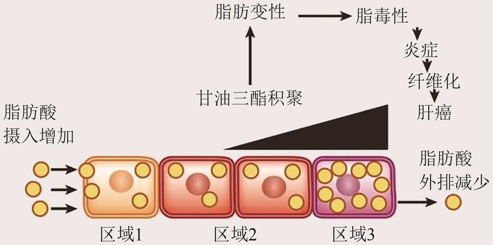
Fig. 3 Spatial imbalance in lipid metabolism can promote the development of NAFLD or NASH
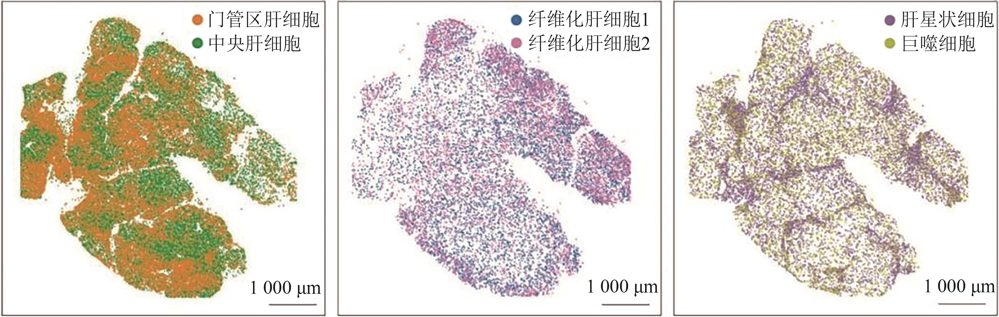
Fig. 4 Spatial transcriptomic profiling of hepatocytes in fibrotic liver[12]

Fig. 5 Cellular interactions and pro-invasive/metastatic mechanisms in the tumor invasive zone
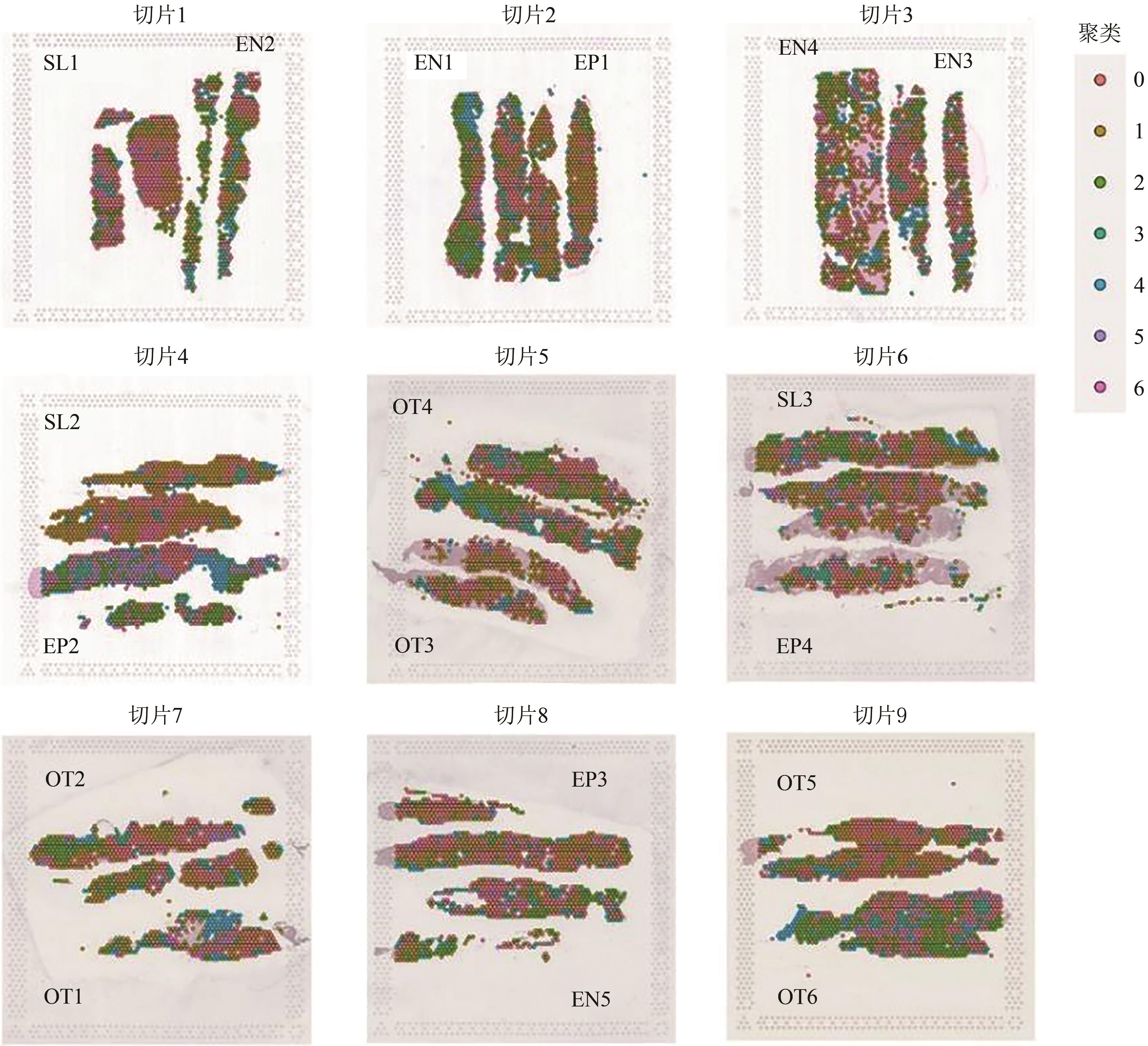
Fig. 6 Spatial heterogeneities in patients with chronic HBV infection
| [1] | MAYNARD K R, COLLADO-TORRES L, WEBER L M, et al.. Transcriptome-scale spatial gene expression in the human dorsolateral prefrontal cortex[J]. Nat. Neurosci., 2021, 24(3): 425-436. |
| [2] | CHEN A, LIAO S, CHENG M, et al.. Spatiotemporal transcriptomic atlas of mouse organogenesis using DNA nanoball-patterned arrays[J]. Cell, 2022, 185(10): 1777-1792. |
| [3] | CHEN K H, BOETTIGER A N, MOFFITT J R, et al.. RNA imaging. Spatially resolved, highly multiplexed RNA profiling in single cells[J/OL]. Science, 2015, 348(6233): aaa6090[2025-06-24]. . |
| [4] | ENG C L, LAWSON M, ZHU Q, et al.. Transcriptome-scale super-resolved imaging in tissues by RNA seqFISH[J]. Nature, 2019, 568(7751): 235-239. |
| [5] | RODRIQUES S G, STICKELS R R, GOEVA A, et al.. Slide-seq: a scalable technology for measuring genome-wide expression at high spatial resolution[J]. Science, 2019, 363(6434): 1463-1467. |
| [6] | WANG X, ALLEN W E, WRIGHT M A, et al.. Three-dimensional intact-tissue sequencing of single-cell transcriptional states[J/OL]. Science, 2018, 361(6400): eaat5691[2025-06-24]. . |
| [7] | BERGMANN S, PENFOLD C A, SLATERY E, et al.. Spatial profiling of early primate gastrulation in utero [J]. Nature, 2022, 609(7925): 136-143. |
| [8] | MO C K, LIU J, CHEN S, et al.. Tumour evolution and microenvironment interactions in 2D and 3D space[J]. Nature, 2024, 634(8036): 1178-1186. |
| [9] | DROIN C, KHOLTEI J E, BAHAR HALPERN K, et al.. Space-time logic of liver gene expression at sub-lobular scale[J]. Nat. Metab., 2021, 3(1): 43-58. |
| [10] | HALPERN K B, SHENHAV R, MATCOVITCH-NATAN O, et al.. Single-cell spatial reconstruction reveals global division of labour in the mammalian liver[J]. Nature, 2017, 542(7641): 352-356. |
| [11] | GEBHARDT R, MATZ-SOJA M. Liver zonation: novel aspects of its regulation and its impact on homeostasis[J]. World J. Gastroenterol., 2014, 20(26): 8491-8504. |
| [12] | WATSON B R, PAUL B, RAHMAN R U, et al.. Spatial transcriptomics of healthy and fibrotic human liver at single-cell resolution[J/OL]. Nat. Commun., 2025, 16(1): 319[2025-06-24]. . |
| [13] | RIAZI K, AZHARI H, CHARETTE J H, et al.. The prevalence and incidence of NAFLD worldwide: a systematic review and meta-analysis[J]. Lancet Gastroenterol. Hepatol., 2022, 7(9): 851-861. |
| [14] | SAVIANO A, HENDERSON N C, BAUMERT T F. Single-cell genomics and spatial transcriptomics: discovery of novel cell states and cellular interactions in liver physiology and disease biology[J]. J. Hepatol., 2020, 73(5): 1219-1230. |
| [15] | MARTINI T, NAEF F, TCHORZ J S. Spatiotemporal metabolic liver zonation and consequences on pathophysiology[J]. Annu. Rev. Pathol., 2023, 18: 439-466. |
| [16] | HUBY T, GAUTIER E L. Immune cell-mediated features of non-alcoholic steatohepatitis[J]. Nat. Rev. Immunol., 2022, 22(7): 429-443. |
| [17] | SCHUSTER S, CABRERA D, ARRESE M, et al.. Triggering and resolution of inflammation in NASH[J]. Nat. Rev. Gastroenterol. Hepatol., 2018, 15(6): 349-364. |
| [18] | PARK E J, LEE J H, YU G Y, et al.. Dietary and genetic obesity promote liver inflammation and tumorigenesis by enhancing IL-6 and TNF expression[J]. Cell, 2010, 140(2): 197-208. |
| [19] | PEISELER M, SCHWABE R, HAMPE J, et al.. Immune mechanisms linking metabolic injury to inflammation and fibrosis in fatty liver disease-novel insights into cellular communication circuits[J]. J. Hepatol., 2022, 77(4): 1136-1160. |
| [20] | RAPPEZ L, STADLER M, TRIANA S, et al.. SpaceM reveals metabolic states of single cells[J]. Nat. Methods, 2021, 18(7): 799-805. |
| [21] | GUILLIAMS M, BONNARDEL J, HAEST B, et al.. Spatial proteogenomics reveals distinct and evolutionarily conserved hepatic macrophage niches[J]. Cell, 2022, 185(2): 379-396. |
| [22] | KISSELEVA T, BRENNER D. Molecular and cellular mechanisms of liver fibrosis and its regression[J]. Nat. Rev. Gastroenterol. Hepatol., 2021, 18(3): 151-166. |
| [23] | TSUCHIDA T, FRIEDMAN S L. Mechanisms of hepatic stellate cell activation[J]. Nat. Rev. Gastroenterol. Hepatol., 2017, 14(7): 397-411. |
| [24] | FRIEDMAN S L. Hepatic stellate cells: protean, multifunctional, and enigmatic cells of the liver[J]. Physiol. Rev., 2008, 88(1): 125-172. |
| [25] | TRIVEDI P, WANG S, FRIEDMAN S L. The power of plasticity-metabolic regulation of hepatic stellate cells[J]. Cell Metab., 2021, 33(2): 242-257. |
| [26] | HAMMERICH L, TACKE F. Hepatic inflammatory responses in liver fibrosis[J]. Nat. Rev. Gastroenterol. Hepatol., 2023, 20(10): 633-646. |
| [27] | CHO C S, XI J, SI Y, et al.. Microscopic examination of spatial transcriptome using Seq-Scope[J]. Cell, 2021, 184(13): 3559-3572. |
| [28] | CHUNG B K, ØGAARD J, REIMS H M, et al.. Spatial transcriptomics identifies enriched gene expression and cell types in human liver fibrosis[J]. Hepatol. Commun., 2022, 6(9): 2538-2550. |
| [29] | MATCHETT K P, WILSON-KANAMORI J R, PORTMAN J R, et al.. Multimodal decoding of human liver regeneration[J]. Nature, 2024, 630(8015): 158-165. |
| [30] | YANG J D, HAINAUT P, GORES G J, et al.. A global view of hepatocellular carcinoma: trends, risk, prevention and management[J]. Nat. Rev. Gastroenterol. Hepatol., 2019, 16(10): 589-604. |
| [31] | WU R, GUO W, QIU X, et al.. Comprehensive analysis of spatial architecture in primary liver cancer[J/OL]. Sci. Adv., 2021, 7(51): eabg3750[2025-06-24]. . |
| [32] | WU L, YAN J, BAI Y, et al.. An invasive zone in human liver cancer identified by Stereo-seq promotes hepatocyte-tumor cell crosstalk, local immunosuppression and tumor progression[J]. Cell Res., 2023, 33(8): 585-603. |
| [33] | XUE R, ZHANG Q, CAO Q, et al.. Liver tumour immune microenvironment subtypes and neutrophil heterogeneity[J]. Nature, 2022, 612(7938): 141-147. |
| [34] | YANG M, SONG X, ZHANG F, et al.. Spatial proteomic landscape of primary and relapsed hepatocellular carcinoma reveals immune escape characteristics in early relapse[J]. Hepatology, 2025, 81(5): 1452-1467. |
| [35] | LI M, WANG L, CONG L, et al.. Spatial proteomics of immune microenvironment in nonalcoholic steatohepatitis-associated hepatocellular carcinoma[J]. Hepatology, 2024, 79(3): 560-574. |
| [36] | LIU Y, XUN Z, MA K, et al.. Identification of a tumour immune barrier in the HCC microenvironment that determines the efficacy of immunotherapy[J]. J. Hepatol., 2023, 78(4): 770-782. |
| [37] | YANG Y, NI Q, LI H, et al.. Genomic and the tumor microenvironment heterogeneity in multifocal hepatocellular carcinoma[J]. Hepatology, 2024. . |
| [38] | GUO D Z, ZHANG X, ZHANG S Q, et al.. Single-cell tumor heterogeneity landscape of hepatocellular carcinoma: unraveling the pro-metastatic subtype and its interaction loop with fibroblasts[J/OL]. Mol. Cancer, 2024, 23(1): 157[2025-06-24]. . |
| [39] | CHENG Y, CHEN X, FENG L, et al.. Stromal architecture and fibroblast subpopulations with opposing effects on outcomes in hepatocellular carcinoma[J/OL]. Cell Discov., 2025, 11(1): 1[2025-06-24]. . |
| [40] | JENG W J, PAPATHEODORIDIS G V, LOK A S F. Hepatitis B[J]. Lancet, 2023, 401(10381): 1039-1052. |
| [41] | PIETSCHMANN T, BROWN R J P. Hepatitis C virus[J]. Trends Microbiol., 2019, 27(4): 379-380. |
| [42] | HSU Y C, HUANG D Q, NGUYEN M H. Global burden of hepatitis B virus: current status, missed opportunities and a call for action[J]. Nat. Rev. Gastroenterol. Hepatol., 2023, 20(8): 524-537. |
| [43] | DE SIMONE G, ANDREATA F, BLERIOT C, et al.. Identification of a Kupffer cell subset capable of reverting the T cell dysfunction induced by hepatocellular priming[J]. Immunity, 2021, 54(9): 2089-2100. |
| [44] | HENSEL N, GU Z, SAGAR, et al.. Memory-like HCV-specific CD8+ T cells retain a molecular scar after cure of chronic HCV infection[J]. Nat. Immunol., 2021, 22(2): 229-239. |
| [45] | SHI J, LI Q, LI J, et al.. Single-cell RNA sequencing reveals the spatial heterogeneity and functional alteration of endothelial cells in chronic hepatitis B infection[J/OL]. Int. J. Mol. Sci., 2024, 25(13): 7016[2025-06-24]. . |
| [46] | YU X, GONG Q, YU D, et al.. Spatial transcriptomics reveals a low extent of transcriptionally active hepatitis B virus integration in patients with HBsAg loss[J]. Gut, 2024, 73(5): 797-809. |
| [47] | CROSS A, HARRIS J M, ARBE-BARNES E, et al.. Characterisation of HBV and co-infection with HDV and HIV through spatial transcriptomics[J/OL]. Gastroenterology, 2024, 2(3): e100067[2025-06-24]. . |
| [1] | Lingfei WAN, Wenting PAN, Yuting YONG, Yuanshuai LI, Yue ZHAO, Xinlong YAN. Research Progress of Single-cell Transcriptome Sequencing Technology in Liver Fibrosis [J]. Current Biotechnology, 2024, 14(5): 793-804. |
| [2] | Shi HUANG, Yinyin MO, Lyujing LUO, Huiting LIU, Zhengyu CHEN, Genliang LI. Bioinformatics Analysis on the Regulation Mechanism of NHP2 on Hepatocellular Carcinoma Senescence [J]. Current Biotechnology, 2024, 14(1): 141-148. |
| [3] | Yanrong WANG, Yuanbiao GUO. Construction of a Prognostic Signature for Hepatocellular Carcinoma Based on Ferroptosis-related LncRNAs [J]. Current Biotechnology, 2023, 13(3): 473-481. |
| [4] | Guanqun LI, Chaojie LANG, Feng XIA. Study on Establishment and Validation of Prognostic Evaluation Model for Hepatocellular Carcinoma Based on Methylomics Database [J]. Current Biotechnology, 2023, 13(2): 282-290. |
| [5] | WANG Jiao-jiao1, GENG Yuan-yuan1, ZHANG Jia-yao2*, JIANG Wei1. Progress for Biomarkers and Detection Methods of Circulating Tumor Cell of Hepatocellular Carcinoma [J]. Curr. Biotech., 2015, 5(2): 113-119. |
| Viewed | ||||||
|
Full text |
|
|||||
|
Abstract |
|
|||||